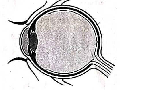
Redraw the diagram. Label it by identifying the parts indicated in the statements given. (a) The part that allows light to enter the eye. (b) The part that adjusts the curvature of the lens. (c) The part where the cone cells are abundant.
Official Solution
Correct Option:
(1)
Step 1: Understanding the eye diagram.
The diagram represents the internal structure of the human eye, where different parts perform specific functions related to vision. Step 2: Part allowing light to enter the eye.
The part that allows light to enter the eye is the pupil. It is an opening present in the center of the iris through which light passes into the eye. Step 3: Part adjusting curvature of the lens.
The ciliary muscles are responsible for adjusting the curvature of the lens. They contract or relax to change the shape of the lens for proper focusing (accommodation). Step 4: Part where cone cells are abundant.
Cone cells are abundant in the yellow spot (fovea) of the retina. This region is responsible for sharp and detailed vision in bright light. Step 5: Conclusion.
Thus, pupil allows light entry, ciliary muscles adjust lens curvature, and fovea contains maximum cone cells for clear vision.
02
PYQ 2026
medium
biologyID: kerala-s
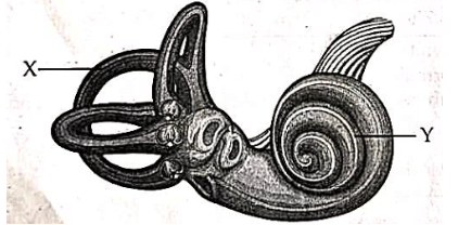
Observe the illustration and answer the following questions. (a) Identify the parts labelled as `X' and `Y'. (b) What will be the effect of the rotational movement of head in `X'? (c) Write the processes in `Y' that leads to hearing.
Official Solution
Correct Option:
(1)
Step 1: Identify the labelled parts.
In the given figure of the inner ear, the part labelled `X' is the semicircular canals and the part labelled `Y' is the cochlea. Step 2: Explain the function of `X'.
The semicircular canals are concerned with maintaining body balance, especially during rotational or angular movements of the head. They contain fluid and sensory hair cells that detect changes in the position and movement of the head. Step 3: State the effect of rotational movement of head in `X'.
When the head rotates, the fluid inside the semicircular canals moves. This movement bends the sensory hair cells, generating nerve impulses which are sent to the brain. As a result, the brain gets information about the direction and speed of rotation, helping to maintain dynamic balance. Step 4: Explain the role of `Y' in hearing.
The cochlea is the hearing part of the inner ear. Sound vibrations reaching the inner ear create waves in the fluid of the cochlea. These waves stimulate the sensory hair cells present in the organ of Corti. Step 5: Describe the process leading to hearing.
The stimulated hair cells convert sound vibrations into electrical nerve impulses. These impulses are then carried by the auditory nerve to the brain, where they are interpreted as sound. Thus, the cochlea helps in the process of hearing.
03
PYQ 2026
medium
biologyID: kerala-s
Identify the fluid indicated by the statements given below and write any two of its functions. • It fills the space between the inner layers of the meninges. • Ependymal cells play a role in the formation of this fluid.
Official Solution
Correct Option:
(1)
Step 1: Understanding the given clues.
The fluid mentioned fills the space between the meninges (protective layers of the brain and spinal cord), and is formed with the help of ependymal cells. Step 2: Identification of the fluid.
The fluid described is Cerebrospinal Fluid (CSF). It is present in the subarachnoid space between the meninges and is produced by specialized ependymal cells in the ventricles of the brain. Step 3: Function 1 – Protection.
CSF acts as a shock absorber, protecting the brain and spinal cord from mechanical injuries by cushioning them. Step 4: Function 2 – Transport and nourishment.
It helps in the transport of nutrients and removal of waste products from the central nervous system, maintaining a stable chemical environment. Step 5: Conclusion.
Thus, cerebrospinal fluid is essential for protection, nourishment, and proper functioning of the brain and spinal cord.
04
PYQ 2026
medium
biologyID: kerala-s
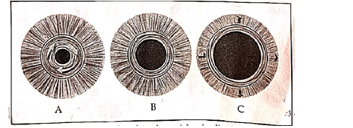
Analyse the figure given and answer the following questions. (a) Identify the figure related to clear vision in dim light. (b) Which muscular activities take place here?
Official Solution
Correct Option:
(1)
Step 1: Understanding the figures.
The given figures (A, B, and C) represent different conditions of the pupil of the human eye under varying light intensities. The size of the pupil changes depending on the amount of light entering the eye. Step 2: Identifying dim light condition.
In dim light, the pupil becomes large (dilated) to allow more light to enter the eye for better vision. Among the given figures, Figure C shows the largest pupil. Step 3: Answer for part (a).
Therefore, the figure related to clear vision in dim light is Figure C. Step 4: Muscular activities involved.
The change in pupil size is controlled by the muscles of the iris:
- Radial muscles contract to dilate the pupil (in dim light).
- Circular muscles relax during this process. Step 5: Conclusion.
Thus, in dim light, radial muscles contract and circular muscles relax, causing pupil dilation to allow more light inside the eye.
05
PYQ 2026
medium
biologyID: kerala-s
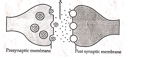
Structure of synapse is given. Analyse it and answer the following questions. (a) Identify the chemical labelled as `X'. (b) Mention their role in the transmission of impulse to only one direction.
Official Solution
Correct Option:
(1)
Step 1: Identify the labelled part `X'.
In the given synapse diagram, the label `X' indicates the chemical substances present in the synaptic cleft. These chemicals are called neurotransmitters. Step 2: Explain what neurotransmitters do.
Neurotransmitters are released from the synaptic vesicles of the presynaptic membrane when a nerve impulse reaches the end of the axon. These chemicals diffuse across the synaptic cleft and carry the signal to the postsynaptic membrane. Step 3: State the answer for part (a).
Therefore, the chemical labelled as `X' is neurotransmitter (for example, acetylcholine in many synapses). Step 4: Explain transmission in one direction.
Transmission of nerve impulse across a synapse takes place only in one direction because neurotransmitters are released only from the presynaptic terminal. The receptors for these neurotransmitters are present only on the postsynaptic membrane. Step 5: Conclude the role in one-way conduction.
As a result, the impulse passes from the presynaptic neuron to the postsynaptic neuron only, and not in the reverse direction. This ensures unidirectional transmission of nerve impulses.
06
PYQ 2026
medium
biologyID: kerala-s
When we feel fear or tension, the heartbeat increases and digestion slows down without our control. (a) Name the division of the autonomic nervous system that controls these actions. (b) Write any two other actions controlled by this nervous system during such situations.
Official Solution
Correct Option:
(1)
Step 1: Understand the situation described.
The condition of fear or tension represents a stress or emergency situation in the body. During such situations, certain involuntary actions like increased heartbeat and reduced digestion occur automatically. Step 2: Identify the responsible division.
These responses are controlled by the sympathetic division of the autonomic nervous system, which prepares the body for fight or flight response. Step 3: Answer part (a).
Therefore, the division responsible is the sympathetic nervous system. Step 4: Identify other actions during stress.
During such situations, the sympathetic nervous system also:
- Dilates the pupils to allow more light into the eyes
- Increases the rate of breathing to supply more oxygen
- Diverts blood flow from digestive organs to muscles
- Causes sweating Step 5: Answer part (b).
Any two actions are: pupil dilation and increase in breathing rate.
07
PYQ 2026
medium
biologyID: kerala-s
Analyse the process of taste detection and choose the correct sequence of steps from the options given: Steps:
(i) Substances dissolve in saliva.
(ii) Nerves carry impulses to brain.
(iii) Chemoreceptors generate impulses.
(iv) Substances reach the minute pores in the papilla.
1
(iii), (ii), (i), (iv)
2
(ii), (i), (iii), (iv)
3
(i), (iv), (iii), (ii)
4
(i), (iii), (ii), (iv)
Official Solution
Correct Option:
(3)
Step 1: Understanding initial process. Taste detection starts when food substances dissolve in saliva. This is necessary because taste receptors can only detect chemicals in dissolved form. Step 2: Movement towards receptors. After dissolving, the substances reach the minute pores present in the papilla (taste buds) on the tongue. These pores allow the dissolved chemicals to interact with sensory cells. Step 3: Generation of impulses. The chemoreceptors present in the taste buds get stimulated by these dissolved substances and generate nerve impulses. Step 4: Transmission to brain. Finally, the nerves carry these impulses to the brain, where the sensation of taste is perceived. Step 5: Conclusion. Thus, the correct sequence is (i) → (iv) → (iii) → (ii).
About Central Nervous System - KERALA-SSLC-EXAM
Central Nervous System is a vital chapter for KERALA-SSLC-EXAM aspirants. Mastering the concepts covered in this chapter is essential for securing a top rank.
By rigorously practicing the previous year questions associated with this chapter, you can identify high-yield topics, understand the examiner's perspective, and boost your confidence during the actual exam.
Frequently Asked Questions
Why focus on Central Nervous System PYQs?
Analyzing PYQs for this specific chapter reveals the most frequently tested concepts and the typical complexity of questions, allowing you to tailor your study plan efficiently.
How to best use this analysis?
Review the topic breakdown to see which sub-topics within Central Nervous System carry the most weight. Then, tackle the questions iteratively to solidify your understanding.