Lateral ventricles of cerebral hemispheres communicate with third ventricle through __________.
1
foramen ovale
2
foramen of Monro
3
foramen magnum
4
hypophyseal fossa
Official Solution
Correct Option:
(2)
Step 1: Understanding ventricular system. The brain contains a series of interconnected cavities called ventricles that circulate cerebrospinal fluid. Step 2: Connection between ventricles. Each lateral ventricle communicates with the third ventricle through an opening known as the interventricular foramen or foramen of Monro. Step 3: Conclusion. Hence, the correct answer is (B) foramen of Monro.
02
PYQ 2020
medium
biologyID: mht-cet-
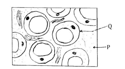
In the above diagram, what do the P and Q indicate?
1
P - Medulla; Q - Macrophage
2
P - Cytoplasm; Q - Fibroblast
3
P - Matrix; Q - Mast cell
4
P - Matrix; Q - Adipocyte
Official Solution
Correct Option:
(4)
Step 1: Understanding the diagram. The diagram represents a tissue section with various types of cells. In such diagrams, the P and Q indicate different cellular structures. Step 2: Analyzing the options. (A) P - Medulla; Q - Macrophage: Incorrect — This does not match the appearance of the cells shown in the diagram. (B) P - Cytoplasm; Q - Fibroblast: Incorrect — The diagram does not indicate cytoplasm and fibroblasts clearly. (C) P - Matrix; Q - Mast cell: Incorrect — This does not match the appearance of the tissue cells. (D) P - Matrix; Q - Adipocyte: Correct — The cells are identified correctly based on the structure shown in the diagram. Step 3: Conclusion. The correct answer is (D) P - Matrix; Q - Adipocyte, as the P is the matrix and Q is identified as adipocyte in the diagram.
03
PYQ 2020
easy
biologyID: mht-cet-
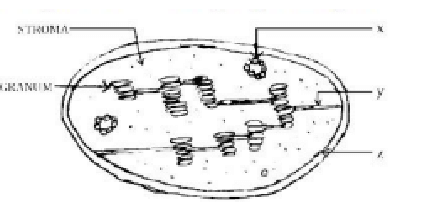
Identify the labels correctly in the given diagram.
Official Solution
Correct Option:
(1)
04
PYQ 2020
medium
biologyID: mht-cet-
The tubular, colourless delicate prolongations of epidermal cells are seen in which region of root in plants?
1
Cell division
2
Absorption
3
Maturation
4
Cell elongation
Official Solution
Correct Option:
(2)
Step 1: Understanding the question. The question asks about the region of the root where the tubular, colorless prolongations of epidermal cells are found. These structures are known as root hairs. Step 2: Analyzing the options. (A) Cell division: The region of cell division is where cells actively divide to form new cells but does not feature root hairs. (B) Absorption: Correct — Root hairs are found in the region of absorption, where they increase the surface area for water and mineral uptake. (C) Maturation: In the maturation region, cells mature and differentiate, but root hairs are mostly absent. (D) Cell elongation: In this region, cells elongate, but root hairs are mainly present in the absorption zone. Step 3: Conclusion. The correct answer is (B) Absorption, as root hairs, which are tubular, colorless projections, are primarily found in this region.
About Anatomy - MHT-CET
Anatomy is a vital chapter for MHT-CET aspirants. Mastering the concepts covered in this chapter is essential for securing a top rank.
By rigorously practicing the previous year questions associated with this chapter, you can identify high-yield topics, understand the examiner's perspective, and boost your confidence during the actual exam.
Frequently Asked Questions
Why focus on Anatomy PYQs?
Analyzing PYQs for this specific chapter reveals the most frequently tested concepts and the typical complexity of questions, allowing you to tailor your study plan efficiently.
How to best use this analysis?
Review the topic breakdown to see which sub-topics within Anatomy carry the most weight. Then, tackle the questions iteratively to solidify your understanding.